

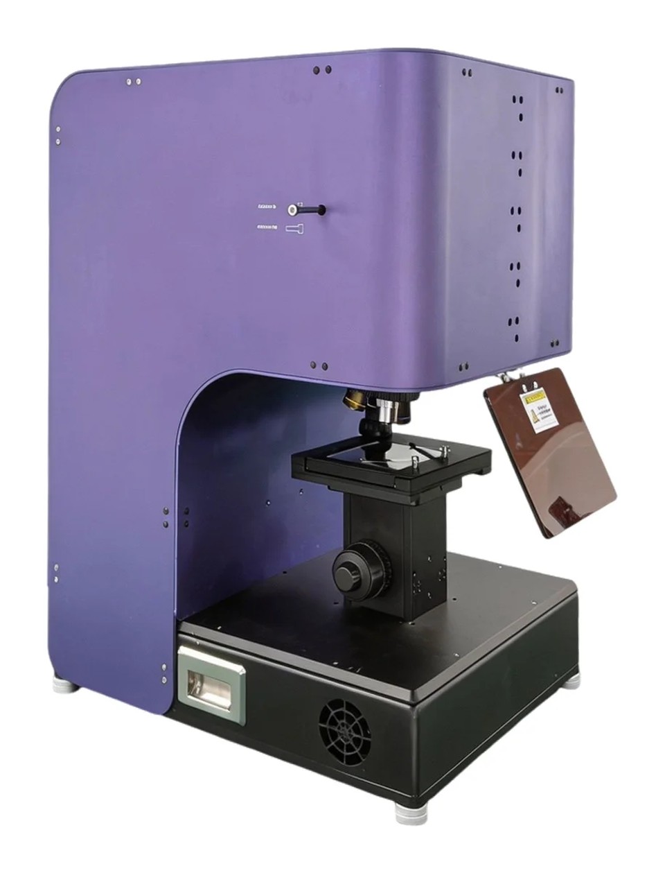
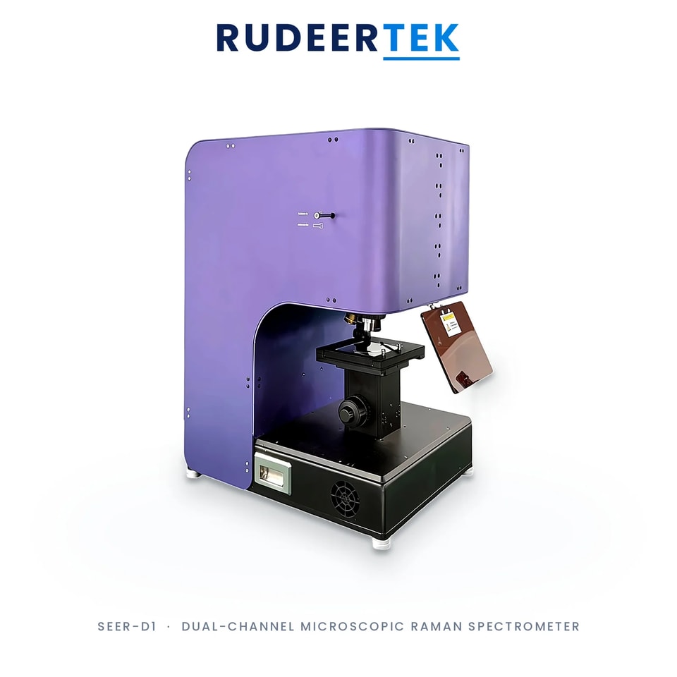
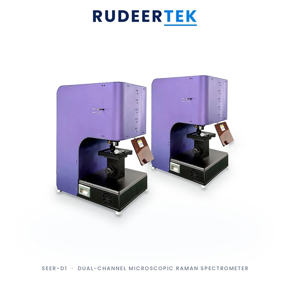
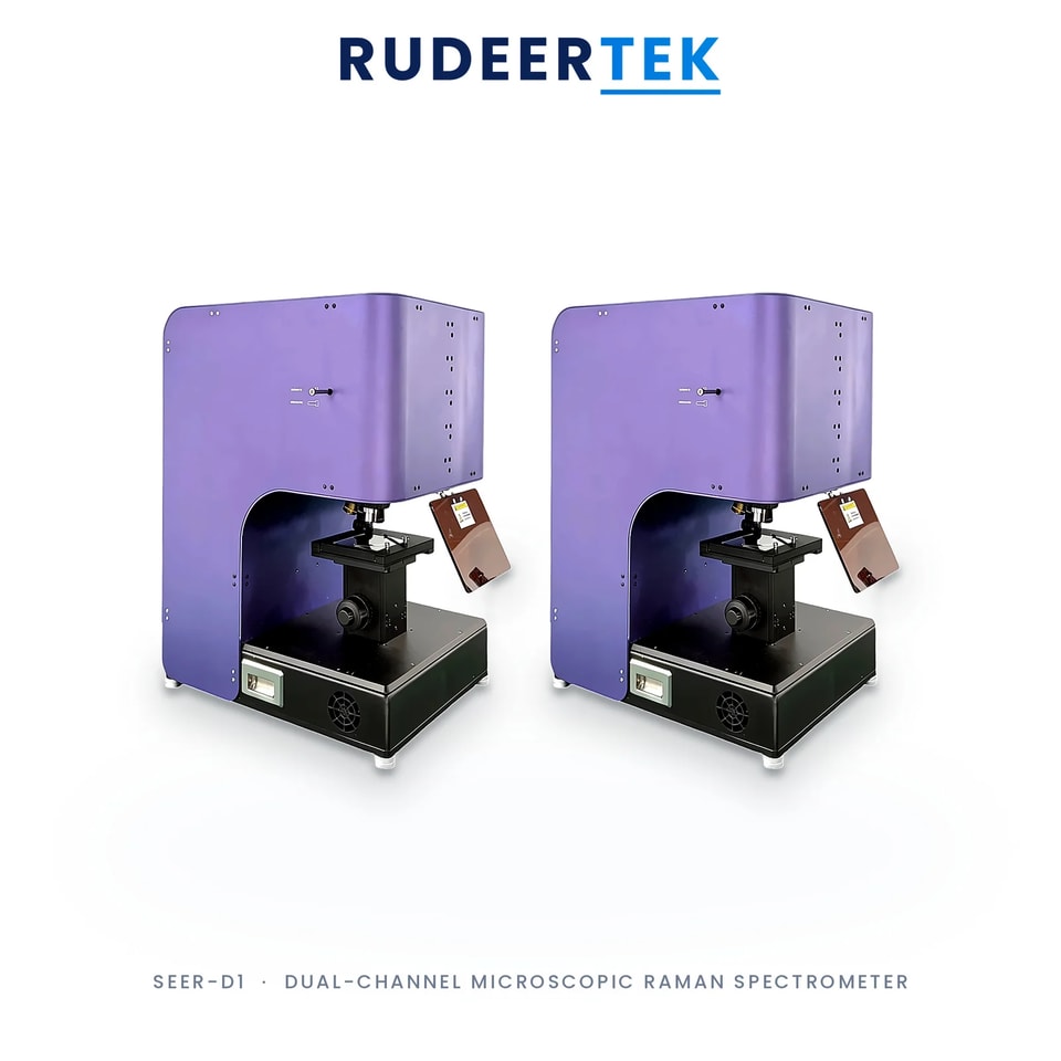

SEER-D1 Dual-Channel Microscopic Raman Spectrometer
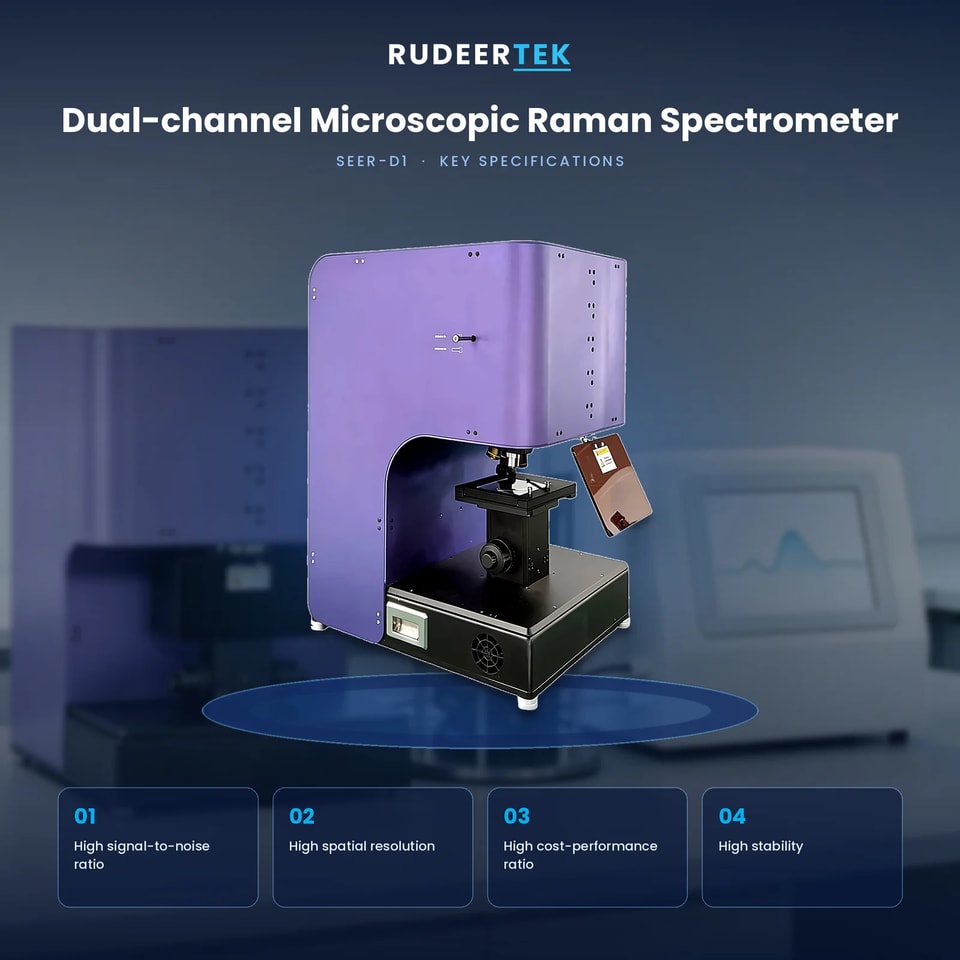
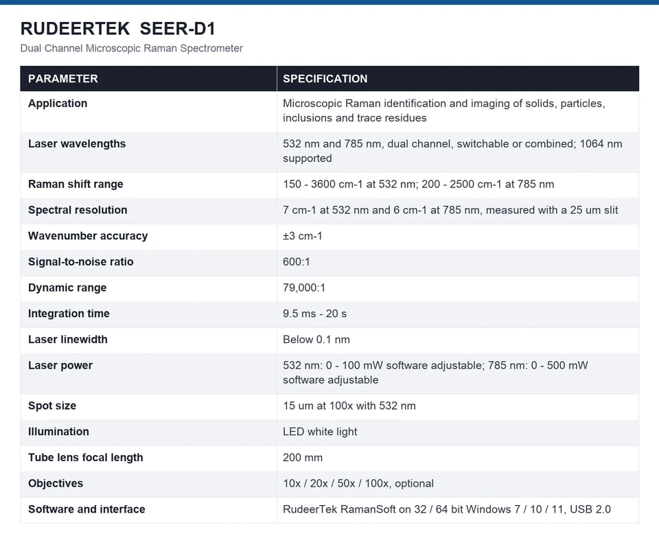
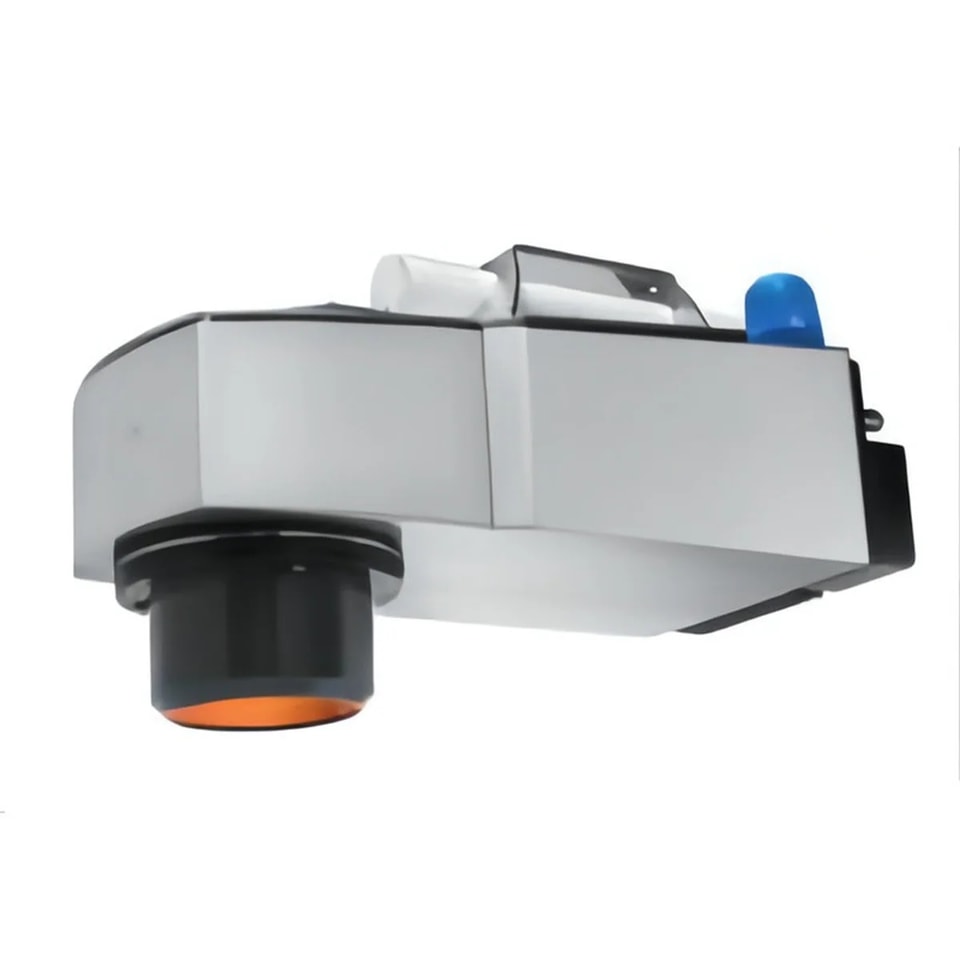
The SEER-D1 is a dual-channel microscopic Raman system: a Raman spectrometer and a microscope share one optical path, so a laser spot of about 15 um at 100x can be placed on a particle, a inclusion or a fibre and the spectrum collected from that exact point. Two laser channels, 532 nm and 785 nm, can be switched or combined under software control, which lets the user take the stronger signal from 532 nm on a benign sample and drop to 785 nm when the sample fluoresces or burns. The Raman range is 150 to 3600 cm-1 at 532 nm and 200 to 2500 cm-1 at 785 nm, with a spectral resolution of 7 and 6 cm-1 respectively at a 25 um slit, wavenumber accuracy of ±3 cm-1, a signal-to-noise ratio of 600:1 and a dynamic range of 79,000:1. Objectives of 10x, 20x, 50x and 100x are available, the laser spot and the microscope image are observed simultaneously, and the whole system runs from RudeerTek RamanSoft on Windows.
· One optical path for microscope and spectrometer, so a 15 um spot at 100x can be measured and identified in place.
· Dual laser channels of 532 nm and 785 nm, switchable or combined in software, to trade signal against fluorescence.
· Raman range 150 - 3600 cm-1 at 532 nm and 200 - 2500 cm-1 at 785 nm, with 7 and 6 cm-1 resolution at a 25 um slit.
· Wavenumber accuracy of ±3 cm-1 and a 79,000:1 dynamic range, so a weak trace band survives beside a strong matrix band.
· Laser spot and microscope image observed at the same time, which is what makes it practical to aim at a single particle.
· Objectives of 10x, 20x, 50x and 100x, LED white illumination, and RudeerTek RamanSoft with calibration functions.
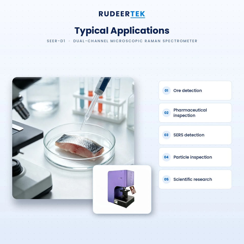
The SEER-D1 is used where a Raman spectrum has to be attributed to a specific microscopic feature: mineral and ore identification with inclusions, pharmaceutical polymorph and counterfeit screening, jadeite and gemstone authentication, SERS trace detection on a substrate, microplastic and particle inspection, forensic trace evidence, and academic research on materials and biological samples.
Which laser should I use, 532 or 785 nm?
532 nm gives a much stronger Raman signal because the intensity scales with the fourth power of frequency, so it is the first choice for inorganic and mineral samples. 785 nm penetrates further and excites less fluorescence, so it is the choice for coloured, organic, biological and polymer samples that burn or glow at 532 nm.
How small a feature can I measure?
With the 100x objective the laser spot is about 15 um at 532 nm, so a single inclusion or a fibre of that size can be measured. The microscope image and the laser spot are observed simultaneously, which is what lets the operator confirm that the spot is on the feature and not beside it.
Is it confocal?
The system images the laser spot through the microscope objective and collects the Raman scattering back through the same path, which gives depth discrimination at the focal plane. The 200 mm tube lens and the objective selection set the working distance and the axial resolution.
What is SERS used for here?
Surface-enhanced Raman scattering, where the sample is placed on a structured metal substrate that amplifies the signal by orders of magnitude. It is what makes trace detection at low concentration possible with a microscope system, and it is one of the documented applications.
How is the instrument calibrated?
The system provides Raman spectrum calibration, and the wavenumber accuracy of ±3 cm-1 is maintained against a standard such as silicon or a calibration lamp. The laser linewidth below 0.1 nm is what keeps the instrument function narrow enough for the resolution figures.
Solutions & Support
We go the extra mile to thoroughly analyze each project and deliver tailored business solutions that meet your specific requirements.
Contact US
×